

Angiology anatomy
📦 Pek 'Angiology anatomy' mengandungi 30 pelekat pelekat mesti ada. Muat turun percuma untuk dipasang di WhatsApp.
-
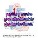
Aorta papillary muscles give attachment to ventricle chordae tendineae. Right ventricle In a normal heart, blood flows from the heart to the body through the aorta.
-
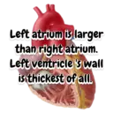
Left atrium is larger than right atrium. Left ventricle's wall is thickest of all.
-
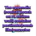
The appendix (conical pouch on rt. atrium wall) contains pectinate(comb ount Sinai Health System like) muscles De-oxygenated blood
-
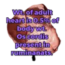
Wt. of adult heart is 0.5% of body wt. Os cordis present in ruminanats.
-

Pericardium is Aubunal aorta attached to phragm <sternum by two pericardico-ster nal ligaments
-
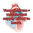
Vasa vasorume vessels that supply blood to heart.
-

moderator band/ septomarginal trebecula- prevent excess dilation of heart.
-
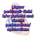
Liquor pericardi-fluid b/w parietal and viceral pericardium/ epicardium
-
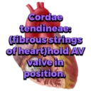
Cordae tendineae: (fibrous strings of heart) hold AV valve in position.
-

5/7 th portion of heart lies on left side because rt. lung is larger in size. Sternum aorta
-
Descending aorta- Pulmonary Circumfle left heart of fowl is Cranial venacava placed at cranial Right atrium part of thoraco- abdominal Right ventricle Left ventricle- cavity
-

Descending aorta- Pulmonary arteries- Pulmonary vein Caudal venacav Circumflex branch of left coronary artery Brachiocephalic trunk Pericardico- phrenic acava Right atrium -Right coronary artery ligament present in dog.
-

The common pulmonary trunk is connected to the descending aorta by fibrous cord called LIGAMENTUM ARTERIOSUM
-

Parts of vessels: tunica intima tunica media tunica adventitia
-
Vasa vasorum: vessels for thick blood vessels; Heart itself is derived from blood vessels
-

Anastomosis- communication b/w blood vessels, 2 types: inter-atrial and arterio-venous
-

End arteries-which do not form any pre-capillary anastomosis.
-

Descending aorta- Pulmonary arteries- Pulmonary veins. Caudal venacava Circumflex left corona Brachiocephalic trunk Cranial venacava In fowl, rt. AV valve is -Right huidm guarded by only one strong muscular leaf. Left ventricle-
-
Red Bond Cott aorta:2 parts ascending-beginning portion; decending caudal aortă
-

aortic arch-sharp curve of ascending aorta, whichpasses caudally and lead to descending aorta
-

aortic sinus/bulbus aorticus:it is the bulbus swelling on the wall of aorta just at the beginning
-

Red Blood Colly brachiocephalic. trunk:large vessel originate from aortic *arch at level of 4th rib and proceed cranially
-

brachiocephalic trunk detaches left and right axillary/subclavian artery
-

In dogs and pigs, left axillary/subclavian... artery arises directly from aortic arch and White Wood Cell not detached from brachiocephalic trunk
-

Close to thoracic inlet and ventral to trachea, the bicarotid trunk bifurcate intort. & left common carotid artery.
-

UMBILICAL ARTERY UMBILICAL VEIN UMBILICAL CORD BLOOD in foetal stage, 2 umbilical arteries and 1 umbilical vein (left BLICAL vein)are present AMNION WHARTON'S JELLY TISSUE
-

UMBILICAL VEIN UMBILICAL CORD BLOOD umbilical vein carry oxygenated blood towards liver. umbilical artery carry UMBILICAL ARTERY deoxygenated blood. WHARTON'S JELLY TISSUE
-

Ductus arteriosus connect pulmonary artery with desc. aorta. After birth, it occluded to form ligamentum arteriosus. e arteries
-

Foramen ovale-connects rt. atrium with left atrium to pass max. blood. Hypogastre arteries
-

umbilical vein join with: 1.portal vein 2 hepatic vein 3.ductus venosus 4.abdominal vena cava 5.rt.atrium
Muat Turun dan Pasang di WhatsApp
Aplikasi Sticker Maker diperlukan untuk memasang pek pelekat ini di WhatsApp anda. Jika anda tidak memilikinya, muat turun dari: Google Play Store dan App Store. Setelah anda memilikinya di telefon anda, ikuti arahan berikut:
- Muat turun fail pelekat yang kami sediakan di bawah (ia mempunyai sambungan .wastickers). Cari fail yang dimuat turun di telefon bimbit anda dan ketuk. Peranti anda akan bertanya aplikasi mana yang ingin anda gunakan untuk membukanya; pilih Sticker Maker. Aplikasi akan secara automatik mengenali fail sebagai pek pelekat baru.
- Setelah anda melihat pek di dalam Sticker Maker, cukup tekan butang yang bertuliskan “Tambah ke Perpustakaan Saya”, kemudian "Tambah ke WhatsApp" dan selesai! Pelekat baru anda akan tersedia untuk digunakan di semua perbualan WhatsApp anda.
Pemasangan di WhatsApp dengan Sticker.ly
Cara mudah untuk menambah pek ini adalah melalui aplikasi Sticker.ly. Jika anda belum memilikinya, muat turun dari: Google Play Store dan App Store. Kemudian pilih salah satu daripada dua kaedah mudah ini:
- Dengan Pautan Langsung: Cuma ketuk pautan ini ke pek PB1H0U. Jika anda memasang Sticker.ly, anda akan segera melihat pelekat dalam aplikasi.
- Menggunakan Kod Pek: Jika anda mahu, anda boleh menyalin "kod pek" dengan mengetuk di sini: dan menampalnya di bar carian di dalam aplikasi Sticker.ly.
Setelah berada di dalam Sticker.ly, sahkan tindakan dengan mengetuk "Tambah ke WhatsApp" dan pelekat akan dihantar serta-merta ke aplikasi WhatsApp anda.


